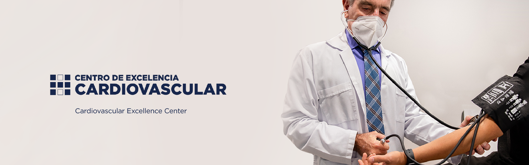
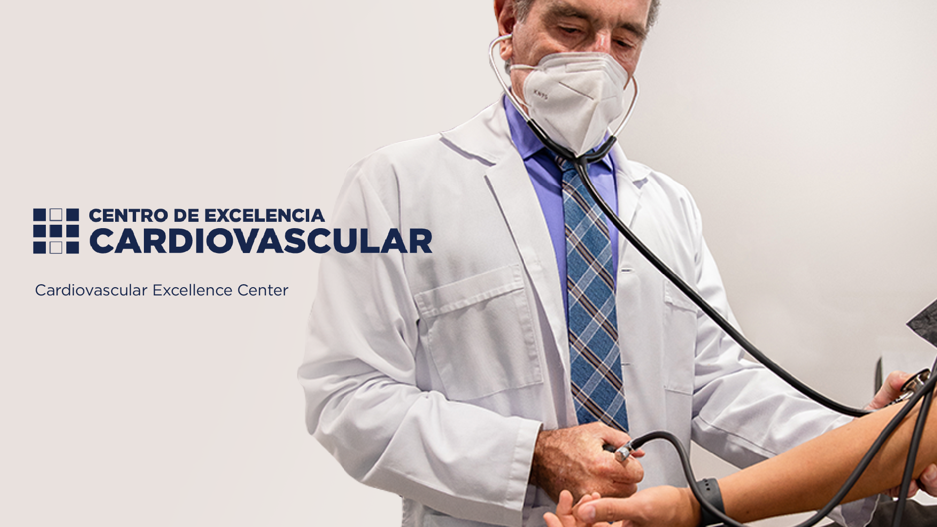
The main objective of the Cardiovascular Center of Excellence at the Metropolitan Hospital is to improve the quality of life of our patients through a new strategy that involves a comprehensive approach to cardiovascular risk factors.
It is a new service model that includes prevention of these risk factors, diagnosis, and specialized treatment for the management of these diseases.
"The main idea of the Center is to be 100% effective in all cardiac diseases, from the simplest to the most complex, such as providing care for open-heart surgery. We want to comprehensively cover all these cardiovascular pathologies from prevention to the most complex resolution procedures," says Dr. Mauricio Obón Dent, Director of the Cardiovascular Center of Excellence at the Metropolitan Hospital.
Cardiovascular diseases (CVD) affect the heart, blood vessels, and encompass a wide range of conditions that can cause deterioration of heart function. Additionally, they often present without pain or obvious symptoms, according to Dr. Obón.
Heart failure, hypertension, diabetes, and obesity are some of the most common diseases in patients who come to the Metropolitan Hospital seeking evaluation and treatment. The Cardiovascular Center of Excellence has qualified personnel, state-of-the-art imaging technology, and devices to be able to manage these pathologies.
Finally, the Cardiovascular Center of Excellence has an angio-graph to diagnose and treat diseases of blood vessels. This technological tool allows for the immediate diagnosis of heart problems.
An angiography machine is a device that emits X-rays and allows for the observation of blood vessels and vascular structures. To achieve its goal, a substance called "contrast medium" is used, which when injected into a blood vessel (artery or vein), makes the vascular anatomy and defects visible on a high-resolution screen through computer processing.
The angiography machine is used by specialists in advanced Interventional Cardiology, Vascular Surgery, Neuroradiology, and Interventional Radiology.
As medicine progresses, technological advances allow us to diagnose and treat medical problems more effectively. Many of the pathologies that were previously treated with highly invasive and risky surgeries are now treated more quickly and safely through the use of the angiography machine. By using these types of state-of-the-art equipment, we achieve less invasive, faster, and more precise interventions. Additionally, the recovery after an endovascular procedure is noticeably shorter and less uncomfortable.
An endovascular procedure is a minimally invasive treatment of lesions in blood vessels (arteries or veins) through catheters and other elements that navigate the interior of these.
Among the elements used to diagnose our patients and subsequently treat them are materials such as catheters, guides, balloons, stents, endoprostheses, among others. Our angiography machine allows for combining all endovascular procedures in all areas of the body.
In the field of cardiology, the angiography machine is mainly used for three areas: diseases of the heart's arteries, structural pathologies, and pathologies of the heart's electrical system.
Diseases of the heart's arteries are by far the most common and are related to heart attacks, angina pectoris, and cholesterol accumulation in the heart's arteries. Most patients who have a heart attack can be taken to the angiography machine to diagnose and treat the problem. Heart attacks are one of the most common causes of death in the world, and it is a widespread problem in Costa Rica. Thus, having this tool available in the hospital is vital.
Now, structural pathologies encompass those problems of the heart's wall or its valves. Currently, many of these problems can be solved with these less invasive methods. Many patients in the past who had to undergo open-heart surgeries now have the option of receiving less invasive therapies, with shorter and more comfortable recoveries. This field has gained more momentum in recent years, and within the institution, we have doctors very familiar with these procedures.
Finally, diseases of the heart's electrical system include all those problems of cardiac arrhythmias and pacemakers. These are addressed almost 100% with the use of the angiography machine. In addition to pacemaker placement, which is a very effective and recognized therapy, a procedure called ablation is also performed, in which a focus causing arrhythmias is entered through a vein and burned. All of these procedures do not involve much pain and have a very high success rate.
Holter Exam
This consists of a device that is connected to the patient and records the electrical activity of the heart for 24 or 48 hours. When the patient is requested to take the exam, the device is placed on them, and they can continue with their normal activities. After 24 or 48 hours, the patient returns with the device to have it removed and for the results to be analyzed.
Stress Test
This is a technique that uses a treadmill or a continuous belt to primarily detect coronary artery disease. The duration of the test depends on the patient's age, physical condition, and the findings we can detect on the stress electrocardiogram or the patient's symptoms. Preparation for the stress test is straightforward and only requires a two-hour fast and some questions about preexisting conditions and medication use. After the test, the patient can resume their normal activities.
Calcium Score
This is an X-ray test using a specialized technique called computed tomography (CT) that shows images of the heart to detect and measure the presence of calcium-containing plaques in the heart's arteries. These plaques (deposits of cholesterol, fat, calcium, and other blood components) can grow and gradually compromise blood flow to the heart muscle.
This test is used to detect whether the patient has a heart disease and to initiate the necessary treatment. Also, plaques (deposits of cholesterol, fat, calcium, and other blood components) form over time and do not show symptoms until they cause a heart attack.
During the exam, the patient is connected to electrodes on the chest to measure cardiac activity. They lie on their back on a table that will slide into a scanner shaped like a tube. They must remain still while the images are taken, a process that can take 10 to 15 minutes.
+506 2521-9595 | info@metropolitanocr.com | All rights reserved © 2023 | Privacy Policy